Cerebrospinal fluid (CSF) is a fluid that cushions the brain and spinal cord. When a leak occurs, the fluid drains through a hole in the dural layer and escapes via the ear, nose, or spinal tissues. An untreated CSF leak drastically increases the risk of meningitis, as it clears a path for bacteria to infiltrate the brain.
Types of Cerebrospinal Fluid (CSF) Leak
Cranial CSF Leak
Often resulting in clear fluid that leaks from the nose or ear, cranial cerebrospinal fluid leaks are caused by a number of factors, including head trauma, tumors, and intracranial pressure. While the most common symptom of a cranial CSF leak is a headache, there are other symptoms that occur.
- Watery discharge out of one ear or sinus
- Hearing loss or recurrent tinnitus
- Strong and unexplained metallic taste
- Meningitis
Spinal CSF Leak
Spinal leaks are a lot more difficult to diagnose due to their spontaneous nature. A spontaneous CSF leak may need to be diagnosed by using complex tests, like a CT scan, MRI, or measuring pressure via a lumbar puncture. Since watery drainage rarely occurs with spinal CSF leaks, it’s crucial to keep an eye out for other symptoms.
- Dizziness and vertigo
- Nausea and vomiting
- Vision and hearing changes
- Light and sound sensitivity
Are Cerebrospinal Fluid Leaks Dangerous
Yes, severe CSF leaks are dangerous, as cerebrospinal fluid cushions your brain. And, without it, your brain can ‘wiggle’ inside your skull, which dramatically increases your risk of major injury and brain damage. In addition to this, CSF leaks indicate a hole in the dura mater, which clears a path for bacteria, meningitis, and other infections.
Treatment for CSF Leaks from Nose and Ears
For a cranial CSF leak, a range of treatment options are available. Conservative treatment may include a lumbar drain, a pressure-relieving shunt, or even bed rest. But, in severe cases, CSF leak repair may require a surgical approach.
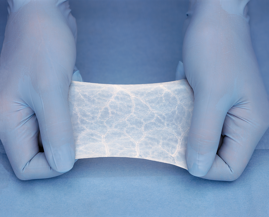
Surgical repair may include endoscopic endonasal repair, an epidural blood patch, or even more invasive measures like open surgery. Everis Medical is dedicated to helping surgeons repair and prevent CSF leaks. Our Biodesign® Duraplasty Graft is designed to completely remodel into the natural host tissue, resulting in a post-op leak rate as low as 1.7%*.
If you’re interested in pursuing easier implantation, stronger seals, and improved outcomes, you should learn more about our Biodesign platform, which is built around the use of an open lattice structure that better allows patient cells to integrate into the dura substitute.
* Bejjani GK, Zabramski J; Durasis Study Group. Safety and efficacy of the porcine small intestinal submucosa dural substitute: results of a prospective multicenter study and literature review. J Neurosurg. 2007;106(6):1028-1033.